Sunday, April 28, 2013

HEPATITIS E - AN EMERGING INFECTIOUS DISEASE

Hepatitis E is listed on the National Institute of Allergy and Infectious Diseases as one of the newly discovered Pathogens in the last twenty years. What is most alarming is that cases are spreading globally. The World Health Organization declares “(e)very year there are 20 million hepatitis E infections, over three million acute cases…and 70 000 hepatitis E-related deaths” (W.H.O.). The virus is caused by drinking water that has been contaminated. Hepatitis E is most often non-life threatening where symptoms in humans disappear after 4-6 weeks. However there are growing numbers of cases that prove to be fatal. Additional new “transmission routes have been identified” (W.H.O.) and this is cause for concern.
 “Hepatitis E is a liver disease caused by the hepatitis E virus: a non-enveloped, positive-sense, single-stranded RNA virus” (W.H.O.). The geographical distribution of Hepatitis E determines the strain of the virus. In poor developing countries like parts of Asia and Africa “genotype 1…is transmitted mainly through the faecal-oral route due to faecal contamination of drinking water” (W.H.O.). Overpopulation, poverty and poor maintenance and “low standards of sanitation increase the risk for transmission of the virus” (W.H.O.). Genotype 3 is most often found in developed countries with the resources and planning for sophisticated sanitation which means this does not spread and cause “community level outbreaks” (W.H.O.).
Water contamination is the cause of most of the hepatitis E outbreaks. Additional outbreaks can occur from “zoonotic transmission from animals to humans…transfusion of infected blood products…(and) vertical transmission from a pregnant woman to her fetus” (W.H.O.). New findings have found that eating raw or improperly cooked shellfish can cause outbreaks in areas susceptible to the virus. Hepatitis E is “no longer confined to Asia and developing countries but has also become a concern of the developed nations” (NCBI).
Because hepatitis E is not contained and new cases are being found globally, this is classified as an emerging infectious disease. If left unchecked it can adversely affect populations of areas where it crops up. Many undeveloped and poor countries already feel the effects of the disease. Many poor people have no access to medical attention, and if they did, they may not be able to afford it. There is scientific proof that diseases and organisms can mutate and change becoming more dangerous. Who’s to say this disease won’t do the same. It could become a wide-spread endemic and wipe out entire cultures. If more and more expectant mothers were infected by Hepatitis E, their newborns would be born with it, and the infant mortality rate would be the cause of a drop in population. The ease of travel could allow this virus to be transported to unaffected areas and migration, along with climatic changes could mutate this organism further.
 
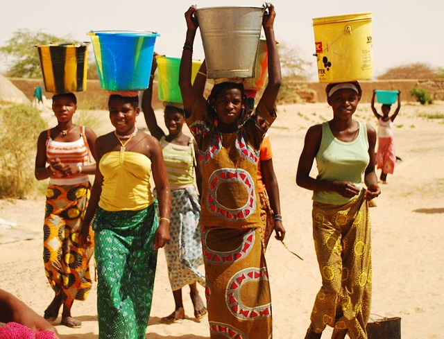
Women walk for miles to carry water back to their families
Photo source: Bing photo images
 How sad that this disease currently affects so many cultures in underdeveloped countries. Because they can’t afford to address the main issue of sanitation, their citizens pay the price. The only thing that could reduce the hepatitis E virus is money. This would provide structure, training and education for “maintaining quality standards for public water supplies; establishing proper disposal systems to eliminate sanitary waste” (W.H.O.). Wouldn’t it be an easy fix to have science come up with a little pill to drop into the water to sanitize it completely, and make it affordable to those countries in need?
Sources:
National Institute of Allergy and Infectious Diseases. US National Library of Medicine. April 03, 2012. Web, 23 April, 2013. http://www.niaid.nih.gov/topics/emerging/pages/list.aspx
World Health Organization. Media Center. Hepatitis E. July, 2012. Web, 23 April, 2013http://www.who.int/mediacentre/factsheets/fs280/en/

Wednesday, April 17, 2013

TECHNOLOGY TO THE RESCUE

I chose to write about surgeries on humans that use innovative products in conjunction with traditional corrective surgery. My research shows that demand for surgeries to repair damaged bone and cartilage has increased. This coupled with profitable investment that encourages successful new discoveries, has led to exciting breakthroughs to enhance surgery. Repair to damaged bone and cartilage in humans interests me. Introducing technology to aid this is a benefit to anyone who lives with pain or damaged and hard-working limbs. Repetitive stress put on our bones and cartilage means that our bones, which contain living cells, may not be able to repair themselves without help. Bones are able to produce more osteoclasts and osteoblasts, to a certain degree, but cartilage often wears thin with time, and this is when humans need additional help.
Watching a video and reading the information below it, ABC news online ran a story about a middle aged man with knee problems due to loss of cartilage. “Cartistem, a stem cell drug used to repair cartilage damage…(is) made from cells found in the umbilical cord blood” (Perez, Sylvia). This new drug combined with “microfracture surgery...is one of the first applications…in the orthopedic space” (Perez, Sylvia). It is being used already in surgeries, but is still in its early stages and requires extensive study and results to back it up further.
Another on-line article showed me a different new technology to aid surgery. “VENADO Foam Strips… (are) designed to enhance bone regeneration and act as an osteogenic stimulus after spine surgery” (Foam Strips). The strips reportedly “are purified fibrillar collagen and biphasic granules… (that) hydrate quickly…and become very flexible and moldable, yet are less likely to dissociate or break apart” (Foam Strips). After surgery when the body is trying to repair the damage on its own, “the implant functions as a scaffold for osteogenic components” (Foam Strips).
In my ignorance of bone surgery, this final piece of research showed a truly interesting approach to “conventional delivery methods of bone graft substitutes during orthopedic surgeries” (Innovative Bone).  By using “preloaded (syringes) with ready-to-use NovaBone Putty…works by delivering an osteoconductive matrix while signaling and stimulating osteoblastic activity to the orthopedic surgical site” (Innovative Bone). This product has the stamp of approval from the FDA and is “a versatile bone graft substitute…(who’s) binder resists migration during irrigation, allowing the bioactive component to stay as placed” (Innovative Bone). How fascinating to think that science developed this product with such a simple delivery approach, using a preloaded syringe to do the task of getting it to the site.
Image: The NovaBone Putty synthetic bone graft
(photo courtesy of NovaBone).

Finally, I was able to research how lucrative technological improvements to standard surgical procedures is. “New analysis from Frost & Sullivan’s …Market research finds the market earned revenue of $1.60 billion in 2012” (Advances). Their findings go on to show that this profit margin will only increase and “the prices for bone graft products have started escalating”(Advances). Not surprisingly, “strong clinical data will give a significant boost to investor’s interest in the bone grafts and bone graft substitutes market” (Advanced).
To summarize, I found that Cartistem, VENADO Foam Strips, and NovaBone Putty are all manmade products scientifically developed as aids used together with corrective surgeries. Their use in surgeries is increasing rapidly. As long as investors can see proven results coupled with research and reliable data, the sky is the limit and technology that works will be used in the future. Humans of today live longer due to improved nutrition, and healthcare. Why shouldn’t such findings be used to improve our physical strength as we age?
Sources
“Advances in Surgical Technologies Boost Demand for Efficient Bone Graft Substitutes, Finds Frost & Sullivan.” Yahoo Finance. March 27, 2013. Web, 10 April, 2013. http://finance.yahoo.com/news/advances-surgical-technologies-boost-demand-115900157.html
“Foam Strips and Bone Graft Systems Biologics.” Today’s Medical Developments. n.d. Web. 10, April 2013. http://www.onlinetmd.com/medical-bone-graft-systems-k2m-040413.aspx
“Innovative Bone Graft Delivery System for Orthopedic Surgery.” HospiMedica International. October 21, 2013. Web, 10 April, 2013. http://www.hospimedica.com/surgical_techniques/articles/294731659/innovative_bone_graft_delivery_system_for_orthopedic_surgery.html
Perez, Sylvia and Christine Tressel. “Innovative stem cell procedure could repair knee damage.” ABC News. April 9, 2013. Web, 10 April, 2013. http://abclocal.go.com/wls/story?section=news/health&id=9059286

Part 2.
1.    What one assignment or activity you performed in this unit are you the most proud of?
I thoroughly enjoyed the Evolution section in Chapter 22 and so I related to the Lab exercise for that chapter. I chose Tenrecs of Madagascar to write about. Because I enjoyed the Chapter, I enjoyed the Lab, especially as I was able to use my imagination instead of just relying on text and factual material.

2.    Why did you enroll in this biology class?
I enrolled in this Biology class because it was a pre-requisite in my General Education section. I chose this as one of my Lab classes because it sounded interesting and I would rather learn about the human body instead of the earth and plants. I might one day need parts of this course if I continue with my thoughts of becoming a Counselor.

3.    Did you feel prepared for this experience when you registered?
No, I knew I would be out of my depth and be learning material I have never learned before. It is my first Biology class ever, and I didn’t do much of the early chapters when I went to school in Africa. I had my 8th grader son and my college student daughter help me go back to basics as there was a lot of material that was completely new to me.

4.    How was your experience different from your expectations?
I had asked around and heard this is a difficult subject with a lot of new material to learn. I knew going into the class that it would be hard, so I was mentally prepared. I was however, constantly overwhelmed with the amount of detailed material that was presented, and it was a struggle to keep up with a full time job, lots of overtime work hours, and being a wife and mother.

5.    What can I do to help you?
Having emails answered in a timely manner really helped when I had questions. There were a few parts of the course where I struggled to understand the concepts, but you really came through for me. I relied heavily on your recorded chapters because your details explained more than just the print version.

Sunday, April 14, 2013

LAB - CHICKEN LEG DISSECTION

Procedure:
1.       Put on gloves and collect your tools.
2.       Thoroughly rinse and dry one chicken leg. Place it in the dissecting tray.
3.       Examine the outside skin tissue. This is the epidermis and the dimples are where feathers emerged. Using scissors and forceps. 
4.       Cut the skin and peel it away from the muscle.
5.       Notice the clear connective tissue that holds the skin to the muscles. As you peel off the skin, you may need to cut away some of this connective tissue. Work slowly and carefully with scissors and forceps until all skin is removed.

6.       Describe how the connective tissue looks and feels. What type of connective tissue is this?
The connective tissue looks white, silky, fatty and a little stretchy like elastic. It is fibrous connective tissue.
Removing the Chicken Skin


7.       Observe, with your naked eye bundles of muscle tissue surrounding the bones. Separate the bundles of muscles by separating them out with your fingers. Begin by inserting your thumb into the muscle of the lower leg. You will need to push forcefully through the shiny lining (called fascia) over the muscle, but it will give way at the natural separations between the muscle bundles. Continue separating the muscle into bundles by forcing your thumb and fingers through the muscle until you are able to distinguish several separate bundles.

a.      Describe the arrangement of the muscle bundles.
The muscles bundles are skeletal muscles tissues. They are long, fibrous groups of cells with connecting fibrous tissue called fascia. The end is joined with the tendon that joins it to the bone.

Separating the muscles


8.       The strong, shiny, white cords, called tendons, hold the muscle to the bones. Some of these tendons will pull away from the bone as you separate the muscle bundles. Use a probe, if needed, to find the tendons of the chicken leg. Using the dissection scissors, cut across the tendons at Line A (Figure 1). Observe the numerous tendons and pull the freed muscles down and away from the bone, as if you were peeling a banana. Careful you don't cut any ligaments that attach bone to bone. Look closely at the ligaments.

Examine the two bones in the lower leg. The large bone (Bone A) is the tibia. The small, toothpick-like bone (Bone B) is the fibula.
a.      What sort of connective tissue are tendons composed of?
Tendons are made of tough, connective tissue which joins bones to muscles.

Finding the Tendons

9.       Remove a single muscle by cutting the tendons and peeling the muscle away from the bone.

a.      What sort of muscle tissue is represented? How do you know?
It is skeletal muscle because it connects to tendons which attach to bone.

b.      Nerves are generally thin, threadlike white strands found between the muscle and the
nearest bone. Look for the nerve in your specimen. Did you find them? ___Yes

c.       What is the physical difference in the tendon of the insertion when compared to the origin?
A tendon is broader where it attaches to the bone at the origin, but it is compact and narrower at insertion where it is attached to moveable bone.

Tendon shapes differ at origin and insertion points


10.       Remove all remaining muscle to expose the bones of the chicken leg.

a.      What is the soft material inside a bone?
The soft material inside a bone is Yellow bone marrow.

b.      Name three specific types of cells present here. Do not break the bone; it is sharp!
Three types of cells present here are dense, compact bone, spongy bone and the central cavity with yellow bone marrow inside it.

c.       Name three functions of bone.
Three functions of bone are support, allows movement and the formation of blood cells.


Looking inside the bone joints


11.       Cut onto the hinge joint by cutting into the top of the covering of the joint from the femur side. It will become apparent that you must remove the knee cap area to expose the menisci and ligaments within. Pull up on the knee cap area and cut through it with the scissors. You will have cut through the bursa, a sac that acts as a shock absorber for the knee joint. These are found in every joint.

12.       Pull the covering back and look into the inside of the joint. You will see more white bands of ligaments holding the bones together. Observe the shiny, white layer covering the ends of the bones is cartilage. It helps the bones slide smoothly when the leg bends.

13.       Bend the specimen at Joint B (Figure 1) and rotate the femur in all directions. Remove the muscle that covers Joint B by cutting parallel to the femur, upward toward the backbone. Remove pink muscle tissue until you see a shiny white sheet of ligament that covers the joint. Present is an exterior ligament that holds the femur in the hip socket

a.      What type of connective tissue composes the ligaments?
Denser fibrous is what ligaments is made up of to attach bone to bone.
Figure 2 is provided in a separate document.
b.      Label Figure 2 with the names of the joints you observed and the motion they make.

c.       On Figure 2, sketch one muscle origin (the name of the bone indicates the insertion) and one muscle insertion you can see in the leg.

Labeling the joints and drawing in a muscle


14.       Dispose of materials as directed.  Make sure to wash your hands, tools, and work station with plenty of soap and water.